
Cavitation surgery
Cavitation Surgery – FDOJ (Fatty Degenerative Osteolysis of the Jawbone Nico Neuralgia-Inducing Cavitational Osteonecrosis)
What Are Cavitations/FDOJ/NICO?

Cavitations, also called FDOJ (Fatty Degenerative Osteolysis of the Jawbone) or NICO (Neuralgia-Inducing Cavitational Osteonecrosis), are areas of dead or dying bone in the jaw where the bone marrow degenerates. These areas:
- Often do not show on standard X-rays
- May involve the trigeminal nerve, causing chronic facial pain, neuropathic pain, or trigeminal neuralgia (AFP/TRN)
- Can remain silent for years, with little to no signs of inflammation, which is why the condition is sometimes misdiagnosed as “idiopathic”
Why Do Cavitations Develop and cause diseases?
Cavitations are caused primarily by reduced blood flow and ischemia in the jawbone:
- Ischemia (poor blood supply) → causes bone marrow degeneration.
- Necrotic adipocytes (dead fat cells) → release proinflammatory chemokines like RANTES/CCL5, which can irritate nerves.
- Fatty degeneration and myxoid changes → further weaken the bone and trigger chronic pain.
Unlike typical infections, FDOJ is a “silent inflammation” — it lacks swelling, redness, or immune cell infiltration, which is why it often goes unnoticed.
How can Chronic Jaw Osteitis/ NICO lesions cause disease? Visit the amazing web site of Dr. J Lechner: http://www.dr-lechner.de/engl
Cytokine and Chemokine Activity
FDOJ is characterized by overexpression of the chemokine RANTES/CCL5:
- Attracts immune cells such as T-cells, NK cells, and eosinophils to the area [20,21].
- Activates nerves, causing hyperexcitability and neuropathic pain [19,28,30].
- Other cytokines, like FGF-2 and IL-1ra, may be slightly elevated, but TNF-α and IL-6 remain normal [15,16].
This explains chronic pain without visible inflammation.
Fatty Degenerative Changes in Dental Cavitations
In some cases, abnormal fatty tissue may develop within areas of the jawbone that fail to heal properly after a tooth extraction or trauma. Instead of healthy bone regeneration, these hollow spaces can become filled with softened, degenerated fatty material, sometimes described as oily or fatty in appearance.
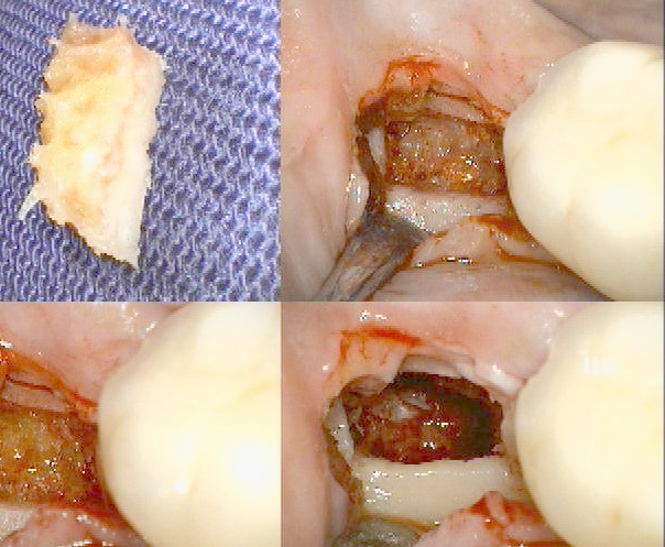
These non-healing defects in the jawbone are commonly referred to as jawbone cavitations. They are associated with compromised bone vitality, reduced blood supply, and ongoing low-grade inflammation. In clinical literature, such conditions may be described using terms such as fatty degenerative osteonecrosis of the jaw (FDOJ) or neuralgia-inducing cavitational osteonecrosis (NICO).
Because the affected bone tissue is no longer healthy, these areas may persist without obvious surface symptoms and can be difficult to detect using conventional dental imaging alone. Proper diagnosis and management often require advanced imaging and clinical assessment.
Pathohistological Findings

Figure 1 – FDOJ sample of fatty and osteolytic degenerated bone marrow (a) and contrast medium X-ray of the FDOJ cavity after curettage (b).
A & B shows a specimen with predominantly fatty transformation of the jawbone (a). The often-impressive extent of FDOJ lesions is illustrated in the right-hand panel by an X-ray with contrast medium.
In 15 patients with AFP/TRN:
| Finding | Number of Patients | % |
|---|---|---|
| AFP/TRN | 15 | 100% |
| Ischemia | 13 | 87% |
| Necrotic adipocytes | 10 | 67% |
| Myxoid degeneration | 12 | 80% |
| Increased fat cells | 12 | 80% |
| Inflammatory cells | 1 | 7% |
Key observations:
- The jawbone shows fatty degeneration and osteolysis, not typical osteomyelitis.
- Inflammatory cells are mostly absent, confirming the “silent” nature of FDOJ.
How Cavitations Cause Pain
- RANTES/CCL5 Overexpression → chronic nerve stimulation and neuropathic pain.
- Neuroplasticity Changes → the brain and spinal cord reorganize in response to constant nerve input, amplifying pain.
- Opioid Receptor Desensitization → RANTES/CCL5 can desensitize μ-opioid receptors, reducing the effectiveness of natural pain suppression and medications
- Peripheral Nerve Sensitization → Schwann cells and immune cells interact with nerves, increasing pain signalling.
Diagnosis
- Standard X-rays often fail to detect FDOJ a CBCT Scan is needed.
- Through-Transmission Alveolar Ultrasonography (TAU) can reliably detect cavitational porosity and is superior to radiograph
Treatment and Outcomes

Figure 3 – Curettage of FDOJ in the lower jaw with denuded infra-alveolar nerve
The clinical example in Figure 3(a) shows the typical situation during surgical debridement and curettage of the lower jaw. The infra-alveolar nerve is totally denuded from its bony sheath by FDOJ. The ischemic process of FDOJ converts the bony sheath, leaving the nerve tissue intact.
Corresponding X-ray without any signs of pathological process in jawbone (b)
As evidenced by what is not shown in the X-ray in the right-hand panel of the figure, this process is inconspicuous and does not show any signs of inflammation or FDOJ. Because of this diagnostic problem of identifying FDOJ on common dental X-rays, this patient suffered from AFP for 7 years and received antidepressants during this time as a singular therapy.
Surgical curettage removes necrotic tissue:
- Reduces RANTES/CCL5 overexpression.
- Provides pain relief in patients with AFP/TRN.
Conclusions
- Cavitations/FDOJ/NICO is hidden jawbone lesions that can cause chronic facial pain.
- Diagnosis requires careful assessment and advanced imaging.
- Surgical curettage is currently the most effective treatment.
- All idiopathic AFP/TRN cases should consider FDOJ/NICO evaluation.
Acknowledgments
Thanks to Professor Dr. GE Bouquot for research on neuralgia-inducing jawbone cavitations.
Disclosure
Dr. Volker von Baehr is co-author. No conflict of interest declared.
References (1-16)
- Jannetta P. J. Arterial compression of the trigeminal nerve at the pons in patients with trigeminal neuralgia. Journal of Neurosurgery. 1967;26(1):159–162. doi: 10.3171/jns.1967.26.1part2.0159. [PubMed] [CrossRef] [Google Scholar]
- Cayley W. E., Jr. Antidepressants for the treatment of neuropathic pain. American Family Physician. 2006;73(11):1933–1934. [PubMed] [Google Scholar]
- Williams C. G., Dellon A. L., Rosson G. D. Management of chronic facial pain. Craniomaxillofacial Trauma & Reconstruction. 2009;2(2):67–76. [PMC free article] [PubMed] [Google Scholar]
- Hargreaves K. M., Cohen S., Berman L. H., editors. Cohen’s Pathways of the Pulp. 10th. St. Louis, Mo, USA: Mosby; 2010. [Google Scholar]
- Evans R. W., Agostoni E. Persistent idiopathic facial pain. Headache. 2006;46(8):1298–1300. doi: 10.1111/j.1526-4610.2006.00541.x. [PubMed] [CrossRef] [Google Scholar]
- Scholz J., Woolf C. J. The neuropathic pain triad: neurons, immune cells and glia. Nature Neuroscience. 2007;10(11):1361–1368. doi: 10.1038/nn1992. [PubMed] [CrossRef] [Google Scholar]
- Tomes J. A course of lectures on dental physiology and surgery. American Journal of Dentistry. 1848;8:p. 209. [Google Scholar]
- Bouquot J. E., Roberts A. M., Person P., Christian J. Neuralgia-inducing cavitational osteonecrosis (NICO): osteomyelitis in 224 jawbone samples from patients with facial neuralgia. Oral Surgery Oral Medicine and Oral Pathology. 1992;73(3):307–320. doi: 10.1016/0030-4220(92)90127-c. [PubMed] [CrossRef] [Google Scholar]
- Mankin H. J. Nontraumatic necrosis of bone (osteonecrosis). The New England Journal of Medicine. 1992;326(22):1473–1479. doi: 10.1056/nejm199205283262206. [PubMed] [CrossRef] [Google Scholar]
- Black R. G. A laboratory model for trigeminal neuralgia. Advances in Neurology. 1974;4:651–658. [Google Scholar]
- Bouquot J., Shankland W., Margolis M. Through-transmission alveolar ultrasonography (TAU)—new technology for evaluation of bone density and desiccation. Oral Surgery, Oral Medicine, Oral Pathology, Oral Radiology, and Endodontology. 2002;93:413–414. [Google Scholar]
- Shankland W. E., II, Bouquot J. E. Focal osteoporotic marrow defect: report of 100 new cases with ultrasonography scans. Cranio. 2004;22(4):314–319. doi: 10.1179/crn.2004.039. [PubMed] [CrossRef] [Google Scholar]
- Lechner J. Validation of dental X-ray by cytokine RANTES—comparison of X-ray findings with cytokine overexpression in jawbone. Clinical, Cosmetic and Investigational Dentistry. 2014;6:71–79. doi: 10.2147/ccide.s69807. [PMC free article] [PubMed] [CrossRef] [Google Scholar]
- Ono K. Symposium: recent advances in avascular necrosis. Clinical Orthopaedics and Related Research. 1992;277:2–138. [Google Scholar]
- Lechner J., von Baehr V. RANTES and fibroblast growth factor 2 in jawbone cavitations: triggers for systemic disease? International Journal of General Medicine. 2013;6:277–290. [PMC free article] [PubMed] [Google Scholar]
- Lechner J., von Baehr V. Hyperactivated signaling pathways of chemokine RANTES/CCL5 in osteopathies of jawbone in breast cancer patients-case report and research. Breast Cancer: Basic and Clinical Research. 2014;8(1):89–96. doi: 10.4137/bcbcr.s15119. [PMC free article] [PubMed] [CrossRef] [Google Scholar]
- an be supplied upon request.